Compact, affordable, low energy
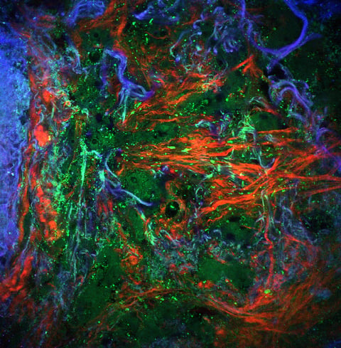
Using 2nd and 3th harmonic generation and 2- or 3-photon excited autofluorescence signals as contrast mechanism, cells, cell nuclei, collagen, elastin and other tissue components, are visualized at an imaging speed up to 2 megapixels per second in 4 modalities.
The technique works at low average powers (< 5 mW), thus avoiding damage inflicted to the fresh tissue sample and allowing it for (re)use in further analysis
An inverted microscope design allows easy positioning of the tissue and makes sure that recording can start immediately, without delay.
Areas of 10×10 mm2 can be scanned within few minutes at a resolution of 0.4 x 0.4 x 2 micrometer in xyz direction.