This proof-of-concept study demonstrates that higher harmonic generation (HHG) microscopy enables rapid, label-free imaging of transbronchial lung cryobiopsies in suspected ILD. On-site within minutes, HHG revealed alveolar structures, fibrosis, and inflammation. This allowed pathologists to assess sample adequacy, and findings were in good concordance with subsequent H&E histology. The results highlight HHG’s potential to reduce sampling errors and speed up diagnostic workflows.
Assessing biopsy adequacy in interstitial lung diseases

This multi-observer study highlights how higher-harmonic generation (HHG) microscopy enables rapid, label-free visualization of fresh pediatric renal tumor specimens. Based on the HHG images pathologists could reliably distinguish non-tumor from tumor tissue with 97% accuracy. The results underscore HHG’s promise as a tool for real-time, intraoperative evaluation and improved sampling strategy in pediatric renal tumor diagnostics.
High diagnostic performances on pediatric renal tumor specimens
This proof-of-concept study shows that higher harmonic generation (HHG) microscopy can rapidly and accurately identify parathyroid tissue during surgery, without staining or freezing. In freshly excised samples, HHG achieved 85% sensitivity and 83% specificity compared with conventional histopathology, while reducing analysis time from around 30 minutes to just a few minutes. The results highlight HHG’s promise as a fast and reliable alternative to frozen sections in endocrine surgery.
Potential alternative to frozen section during thyroid surgery

This multi-observer study explores the use of higher harmonic generation (HHG) microscopy as a fast, label-free imaging method for pediatric gonadal tumor specimens. The results show that HHG can reliably reflect the tissue architecture seen in conventional histology and that pathologists were able to distinguish non-tumoral gonadal tissue from gonadal tumors. These findings support HHG’s promise as a tool for rapid intraoperative evaluation, particularly in evaluating sample representativeness.
On-site histological assessment of pediatric gonadal tumors
This study shows that a portable HHG microscope, combined with an AI-analysis algorithm, can deliver on-site histologic feedback on lung and pleural biopsies in about 6 minutes. Among 109 biopsies from 47 patients, 97% had sufficient image quality for diagnosing malignancy or non-malignancy, and pathologists correctly classified 87% of the HHG images. This work supports HHG + AI as a fast, reliable tool to accelerate diagnosis and guide treatment decisions.
On-site histological feedback on lung and pleural biopsies

This study showcases how third harmonic generation (THG) microscopy, combined with artificial intelligence, can enable real-time, label-free tissue assessment during brain tumor surgery. By instantly distinguishing tumor from healthy tissue, this approach supports faster and more precise intraoperative decision-making. The method achieved high diagnostic performance compared to conventional histology. These results show promising potential for guiding neurosurgeons intraoperatively via immediate feedback on tumor vs non-tumor tissue.
Intraoperative feedback for brain tumors
In diseases such as interstitial lung diseases, immediate feedback on leukocyte ratios has potential to speed-up the diagnostic process and to reduce costs, workload and inter-observer variations. This study shows that label-free THG/MPEF microscopy, paired with a deep learning model, can identify and quantify immune cells (neutrophils, eosinophils, lymphocytes, macrophages) in fresh BAL fluid samples in minutes with >90% accuracy versus standard cytology.
Immune cell differentiation in BAL fluids


Fast and reliable intraoperative pathological feedback using Higher harmonic generation microscopy on unprocessed tissue within minutes, is a promising tool to avoid a two-stage procedure and significantly reduce health care costs in patients undergoing a diagnostic hemithyroidectomy. Images generated show relevant morphological thyroid structures in good accordance with the histology images.
Avoid two-staged thyroid cancer surgery
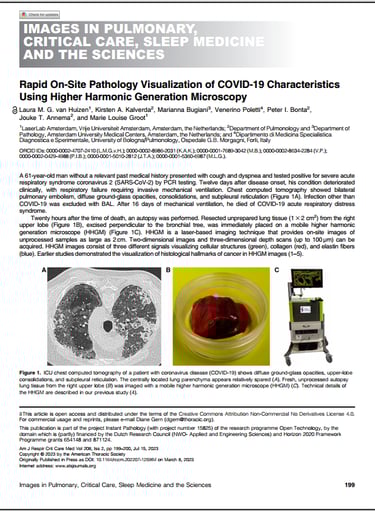
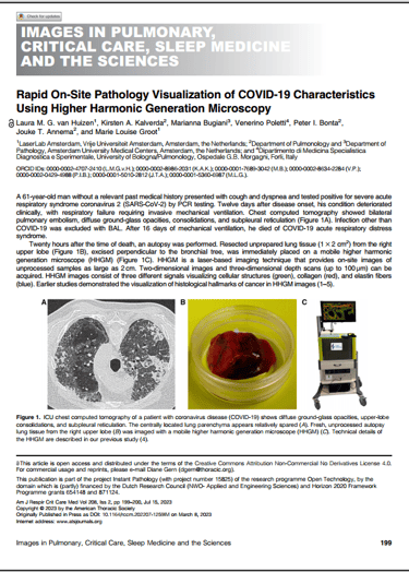
Using higher harmonic generation (HHG) microscopy, the authors enabled real-time, label-free imaging of lung tissue affected by COVID-19. Without any staining or preparation, HHG revealed key pathological features—such as inflammation, fibrosis, and vascular changes with high fidelity to standard histology. The work highlights HHG’s potential for rapid, on-site tissue assessment in infectious and inflammatory lung disease.
On-site pathology visualization of COVID-19 characteristics
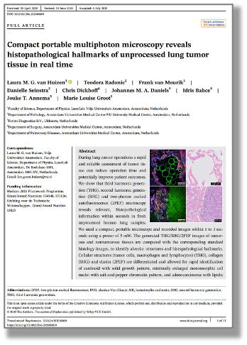
Rapid and reliable histology assessment of tumor tissue can reduce operation time and potentially improve patient outcomes in lung cancer operations. THG/SHG/2PEF imaging in seconds of fresh, unprocesessed human lung tissue is a promising tool for clinical intraoperative assessment of lung tumor tissue. In this study, this digital histology is compared with standard FFPE HE stained histology images.
Showing histological hallmarks in lung tumor resections

Flash Pathology B.V.
Paasheuvelweg 3
1105 BE Amsterdam
The Netherlands
Contact
Menu
Technology
At Flash Pathology, we are committed to empowering you with advanced tools to visualize and understand the intricate details of tissue samples.
Our products are designed and developed under ISO 13485 certified processes